2024 ASGCT Poster Presentation
Non-Viral Cell Engineering of Peripheral Blood Natural Killer Cells Using a GMP-Compliant, Scalable Electroporation Platform
American Society of Gene and Cell Therapy annual meeting
Baltimore, Maryland, USA
May 8, 2024
Our MaxCyte scientist, Lauren Unsworth, presents her poster from the 2024 American Society of Gene and Cell Therapy annual meeting
Natural killer (NK) cells are an integral part of the innate immune system as they recognize an abundance of surface receptors on target cells without the need for antigen specificity and recruit and activate other immune cells by secreting a myriad of cytokines. The hallmark of NK cells is their cytotoxic function which enables them to recognize and attack tumor or infected cells by releasing granules that induce apoptosis of target cells. Electroporation (EP) is a nonviral method for cellular engineering that has been used with virtually all cell types and has been specifically shown to be an effective cell engineering method for peripheral blood NK cells. Advantages of electroporation include highly efficient transfection with a variety of molecules (e.g., DNA, RNA, and ribonucleoprotein (RNP) complexes), regulatory acceptance, and cGMP compatibility. In this study, we optimized conditions for electroporating primary NK cells with the GMP-compliant MaxCyte ExPERT GTx electroporation platform, with specific focus on maximizing cell viability, cell recovery, transfection efficiency, cell expansion and functionality.
Key takeaways
- Previous studies showed that expansion of NK cells before electroporation was necessary for effective transfection
- Electroporation with mRNA had over 90% gene expression and viability
- Electroporation with minicircle DNA (mcDNA) had over 60% expression and viability
- Minimal impact on cell viability, cell expansion, surface marker expression and killing capacity was observed after electroporation
Watch Lauren's poster presentation

View the complete poster that established an optimized process for nonviral cell engineering of primary NK cells using the MaxCyte ExPERT GTx.
Have more questions?
Send your question to one of our cell engineering experts.
Presenter

Popular resources
Poster presentation transcript
Hello everyone., my name is Lauren Unsworth, and today I will be presenting on non-viral cell engineering of peripheral blood natural killer cells using a GMP-compliant scalable electroporation platform.
Natural killer (NK) cells are an integral part of the innate immune system as they recognize an abundance of surface receptors and target cells without the need for antigen specificity and recruit and activate other immune cells by secreting a myriad of cytokines. The hallmark of NK cells is their cytotoxic function, which enables them to recognize and attack tumor or infected cells by releasing granules that induce apoptosis of target cells.
Electroporation, or EP, is a non-viral method for cellular engineering that has been used with virtually all cell types and has been specifically shown to be an effective cell engineering method for peripheral blood NK cells. Advantages of electroporation include highly efficient transfection with a variety of molecules such as DNA, RNA, and proteins as well as regulatory acceptance and cGMP compatibility.
In this study, we optimized conditions for electroporating primary NK cells with the GMP compliant MaxCyte ExPERT GTx electroporation platform with specific focus on maximizing cell viability, cell recovery, transfection efficiency, cell expansion and functionality. Previous studies have shown that expansion of NK cells before electroporation is necessary for effective transfection, and therefore larger scale platforms are required, leading to the use of more costly reagents.
Here, we show that the MaxCyte ExPERT GTx could genetically modify naive or expanded primary NK cells using mRNA, RNP complexes or plasmid DNA. At both a small and larger scale, we observed over 90% gene expression and viability following mRNA electroporation and over 80% expression and viability after minicircle DNA, both at the large and small scale. We also evaluated the effect of commercially available DNA sensing pathway inhibitors to determine if transfection efficiency and cell viability could be increased when loading both small and large plasmid DNA. Furthermore, we knocked out the NKG2A inhibitory receptor at different time points during expansion by loading a single guide RNA, Cas9 RNP complex and looked at post electroporation effects. With our optimized transfection conditions specific for NK cells, we determined that electroporation had minimal impact on cell viability expansion and surface marker expression and killing capacity.
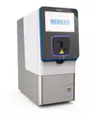
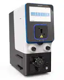
Here, we have a schematic timeline of our non-viral natural killer cell engineering. Peripheral blood mononuclear cells, or PBMCs, were isolated from healthy donor leukopacks. NK cells were isolated using magnetic bead separation and expanded for seven days using a feeder-free culture system. Cells were harvested and electroporated using the GMP-compliant MaxCyte ExPERT GTx electroporation platform and cultured for downstream analysis.
Expanded NK cells were resuspended to 100 million cells per mL in MaxCyte electroporation buffer and mixed with either low- or high-cargo concentrations. Three different cargos were loaded: mRNA, small plasmid DNA or minicircle DNA, noted here as mcDNA, and large plasmid DNA, or pDNA, all expressing GFP. Cells were electroporated with three different preloaded protocols ranging in total energy from low to medium to high. Using the OC-25x3 processing assembly, 25uL of each sample was electroporated and rested for at least 20 minutes at 37°C and subsequently cultured and complete media. Cells were collected at 24, 48 and 72 hours post electroporation for flow cytometry analysis.
We found that a low energy EP protocol and high concentration was optimal for RNA transfection, where a medium energy and high concentration was optimal for both minicircle and large plasmid DNA. With these optimized conditions, GFP expression was achieved at over 90% with mRNA, 80% with minicircle DNA and 40% with large plasmid DNA. Here, we have representative flow cytometry plots of transfection efficiency for each preloaded protocol and every concentration at each time point to post electroporation for mRNA, highlighting our optimal condition of 100 micrograms per mL mRNA and a low-energy protocol. For minicircle DNA or small plasmid DNA, here we're highlighting the optimal condition of 200 micrograms per mL with a medium-energy protocol.
And again, for the large plasmid DNA, highlighting the same optimal condition of 200 micrograms per mL with a medium energy. We then looked at the effect of using inhibitors of the cGAS-STING DNA sensing pathway to see if inhibition would increase DNA toxicity. Four commercially available inhibitors targeting various parts of the pathway were evaluated to determine if inhibition after electroporation could increase transfection efficiency and viability.
Expanded NK cells were electroporated with either no cargo, as a control, or with small minicircle DNA expressing either GFP or CD19-CAR at two different concentrations. A preloaded protocol with medium energy was used, and each sample was rested post electroporation for 20 minutes at 37°C. Immediately following resting, culture media containing either 0.5 micromolar or 1.0 micromolar of each of the four inhibitors were incubated with cells for 20 hours then removed and replaced with fresh media. We found that BX795, MRT67307 and Ru.521 were toxic to the cells.
However, PF06928215 showed to be nontoxic and has the potential to increase transfection efficiency of both GFP and CD19-CAR minicircle DNA expression. However, further analysis and titration is needed. We then sought to assess the ability of the MaxCyte GTx to genetically modify naive or expanded primary NK cells using RNP complexes. NK cells were isolated from PBMCs and either expanded or immediately electroporated using a preloaded protocol with low energy. Single guide RNA targeting the NKG2A locus was mixed with Cas9 at a 1 to 2 ratio and, using the OC-25x3 processing assembly, 25uL of each sample was electroporated using a low-energy protocol and rested for at least 20 minutes at 37°C.
Cells were collected at day three and day seven post electroporation for flow cytometry analysis of viability and NKG2A expression, which the total knockout was calculated from. Results indicate that naive NK cells show similar knockout to NK cells expanded for 4, 7 or 14 days before electroporation, and, therefore, the MaxCyte GTx can efficiently genetically modify NK cells at both a small and larger total cell number.
To determine if electroporation had any impact on NK cell function, expanded NK cells were electroporated with two preloaded protocols with ranging total energies with either mRNA or minicircle DNA. Phenotypic analysis was conducted by flow cytometry, looking at activation markers such as CD16, CD69 and NKp46, and NKG2D, inhibition markers such as NKG2A and maturation marker such as Tim-3.
Cytotoxicity of electroporated NK cells was measured by nonspecific cell targeting of Ramos cells, a B cell lymphoma cell line. Effector to target ratios of NK cells to Ramos cells ranged from 0.25:1 to 2:1. Cells from the samples in the cytotoxicity assay were collected and permeabilized for intracellular staining of Perforin and Granzyme B. The supernatant for each sample in the cytotoxicity assay was harvested, and the concentration of secreted interferon-gamma was measured by ELISA. Each of these assays indicated electroporation had minimal impact on cell functionality, including surface marker expression, cytotoxicity and secretion.
Now that we had optimal electroporation conditions that did not impact NK cell functionality, we scaled up our process using a CD19-CAR mRNA. Here, two different cell numbers were electroporated in two different processing assemblies. The first was the OC25x3, where we electroporated 1 million cells in 25uL. The second was the R20k, where we electroporated 100 million cells in 5mL.
We found that both the small- and large-scale viability was over 80% and transfection efficiency was over 90%, proving our conditions could be efficiently scaled up. Here we had the representative flow cytometry plots of the transfection efficiencies at both scales at 24 hours post electroporation.
To summarize, we were able to establish an optimized process for non-viral cellular engineering of primary NK cells using the Maxcyte ExPERT GTx. DNA sensing pathway inhibitors have the potential to increase the viability and transfection efficiency when electroporated plasmid DNA; however, more testing is needed. The inhibitory receptor, NKG2A, can be efficiently knocked out in naive or expanded primary NK cells. We also found that electroporation had minimal impact on cell viability, surface marker expression, functionality and cytotoxicity, including secretion. We were also able to scale our process up from 1 million cells in 25uL to 100 million cells in 5mL. Thank you for listening.
