Advancements in cell therapy: development of a non-viral gene delivery platform for CAR-T manufacturing

Listen to this webinar hosted by Cell and Gene Therapy Insights about the latest advancements in cell therapy, specifically using homology-independent targeted insertion (HITI) of a therapeutically relevant GD2-CAR transgene into the T cell receptor alpha constant (TRAC) locus using nanoplasmid DNA and CRISPR-Cas9 in primary human T cells.

The lab of Steven Feldman, PhD, is pioneering non-viral gene delivery methods, which promise to mitigate the limitations of viral vectors.

Cell and gene therapy insights logo

Watch the webinar

Slide from presentation of Advancements in cell therapy: development of a non-viral gene delivery platform for CAR-T manufacturing

Challenges with viral vectors

Viral vectors have been the standard in CAR-T cell manufacturing but present several challenges:

  • High costs: establishing manufacturing suites following current good manufacturing practices (cGMP) or outsourcing virus production is financially burdensome
  • Safety issues: viral vectors can induce immunogenic responses and pose risks associated with random genomic integration
  • Regulatory complexities: meeting regulatory standards requires intricate vector production strategies

Non-viral gene delivery—a scalable, replicable process

The lab at Stanford is pioneering non-viral gene delivery methods, which do not have the same limitations or risks of viral vectors. The team uses electroporation to deliver a CRISPR ribonucleoprotein (RNP) and nanoplasmid DNA to edit the cells through homology-independent targeted integration (HITI). This mechanism of action utilizes CRISPR-Cas9 to create a double-stranded breaks in specific loci, facilitating gene insertion without viral vectors. With minimal off-target effects and high yield HITI achieves functional CAR-T cells with comparable functionality to virally transduced cells in preclinical models.

Role of electroporation in non-viral gene delivery
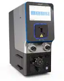
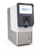
A significant aspect of Steven's work involves the use of electroporation, particularly leveraging MaxCyte's electroporator instruments. Electroporation is a transfection method that uses electrical pulses to introduce gene-editing machinery and other molecules into cells, and MaxCyte's scalable, consistent technology has been an important, but straightforward, step in this process. Using electroporation has the following benefits:

  • Scalability throughout therapy development: MaxCyte's instrumentation can scale up without changing the protocol conditions. This technology can transfect billions of cells in a single run, ensuring process standardization and consistency.
  • Delivery optimization: MaxCyte provides optimized electroporation protocols for a wide range of cell types and payloads, yielding reproducibly high levels of transfection efficiencies and gene editing.
  • Technical support: MaxCyte's cell engineering experts provide extensive technical and scientific support not only for the electroporation process but for the entire cell therapy development workflow, ensuring success for teams developing their cell therapy.
Presenters

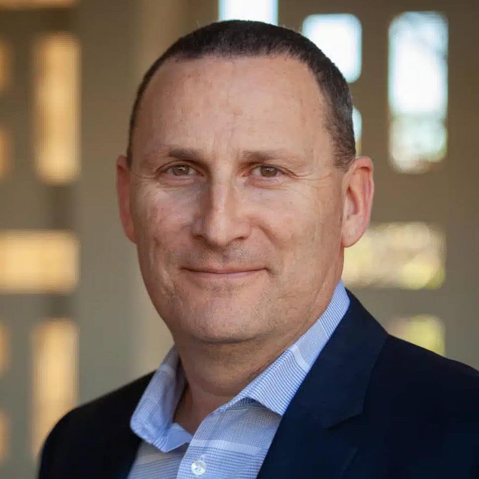
Headshot of Steven Feldman, PhD

Steven Feldman, PhD

Site head and scientific director of the Laboratory for Cell & Gene Medicine at Stanford Center for Cancer Cell Therapy

Steven is a pivotal member of the Stanford Center for Cancer Cell Therapy. He serves as the site head and scientific director of Stanford’s GMP facility and leads a team that focuses on the development and manufacture of novel cell therapies for the treatment of cancer, emphasizing more efficient, cost-effective, and safer gene delivery methods, especially testing other methods than traditional viral vectors.

James-Brady

James Brady, PhD

Senior vice president, technical applications and customer support at MaxCyte

Jim is an experienced biotechnology industry professional with expertise in cell and gene therapy, biologics, and drug discovery. Previously, he was a senior scientist at Genetic Therapy, a Novartis subsidiary, where he worked on lentiviral-based gene therapy treatments and was a group leader at MetaMorphix, managing the company’s transgenic and genetic research programs.

Jim earned a Master of Business Administration degree in finance from The Johns Hopkins University. He completed his postdoctoral fellowship at the National Eye Institute of the National Institutes of Health in Bethesda, Maryland, after obtaining a PhD in genetics from Indiana University in Bloomington, Indiana.

Headshot of Daniel Nguyen

Daniel Nguyen

Director of global sales development and inside sales at MaxCyte

Daniel has extensive experience supporting industry and academic centers with their cell engineering needs from basic research to manufacturing. He has spent the last four years focused on supporting scientists in the areas of cell therapy and CRISPR engineering at MaxCyte.

Transcript

Lauren Coyle: A very warm welcome to today’s Cell and Gene Therapy Insights webinar titled, “Development of a non-viral gene delivery platform for CAR-T manufacturing.” I’m Lauren Coyle, an editor at Bio Insights. And, joining me today are Daniel Nguyen, Jim Brady and Steven Feldman, who will discuss a novel non-viral gene delivery method for CAR-T-cell manufacturing to combat both low-frequency yields and financial burden in the current economic climate. After the presentation, we’ll have a live Q&A session, so feel free to pose your questions to our speakers using the ask a question box at the bottom of your screen, and we’ll try to get to them during the session. Now, I’d like to introduce our guest moderator for today. Daniel’s the director of global sales enablement and inside sales at MaxCyte. Daniel has extensive experience supporting industry and academic centers with their cell engineering needs from basic research to manufacturing. He has spent the last four years focused on supporting scientists in the area of cell therapy and CRISPR engineering at MaxCyte. So, without further ado, I’ll hand over to Daniel who’s going to kick us off and introduce our speakers.

Daniel Nguyen: Thanks, Lauren for the kind introduction. We greatly appreciate everyone for joining us. I’m thrilled to introduce our two speakers for today, Dr. Steve Feldman and Dr. James Brady. Dr. Feldman is the site head and scientific director of Stanford’s GMP facility. He’s also part of the team that leads the Stanford Center for Cancer Cell Therapy. His core team focuses on the development and manufacture of novel cell therapies at Stanford.

Dr. Brady is a senior vice president of technical applications and customer support at MaxCyte. He’s led MaxCyte’s technical teams for nearly 20 years and has a great deal of experience in the cell and gene therapy space. As for the format of the webinar, Dr. Brady will be presenting first and then followed by Dr. Feldman. We’ll wrap up the webinar with a Q&A session. Dr. Brady, I’ll hand things over to you.

James Brady: Okay, thank you, Daniel. So before handing over the presentation to Dr. Felman, I’d just like to take a few minutes to introduce you to MaxCyte’s non-viral GMP-compliant electroporation technology. And I’ll describe some of the strategies that our partners and customers are using to generate CAR-T cells and other types of cellular therapies without the need for viral vectors. Now, viral vectors are widely used in CAR-T manufacturing because historically there were few other options for efficient delivery of genes into primary cells, particularly cells of hematopoietic origin. However, therapeutic manufacturing strategies that rely on viral vectors face a number of challenges. These challenges include high costs associated with either building out a GMP manufacturing suite for large-scale vector production or outsourcing virus production to a CMO. In addition, some viruses trigger immunogenic responses for viral vectors derived from lenti or retrovirus. The risk of producing replication competent virus has always been a concern to regulatory authorities, and mitigating that risk requires complex vector production strategies, often involving division of vector components across multiple plasmids. And in addition, analytical procedure, or procedures, are also recommended to test for replication competent virus. And then finally, with both retroviral and lentiviral gene delivery, there is a risk of the vector integrating into an oncogene or in other undesirable genomic locus. And this risk was recently highlighted in guidance, in a guidance document from the FDA.

Now, MaxCyte was founded 25 years ago to enable non-viral engineering of primary cells for therapeutic applications. Our proprietary flow electroporation technology was developed specifically for use in the clinic. And our mission from the very beginning has been to enable highly efficient loading of molecules into precious cells from patients while imparting minimal impact to cell viability. We offer several different instruments, or instrument models rather, to support researchers and clinicians at all phases of the therapeutic discovery and translational spectrum. Our technology is employed by academic researchers, large pharma, biotech, as well as CROs, CMOs, and CDMOs for many applications including cell therapy, but also for bioproduction and drug discovery.

So, the flow electroporation technology developed by MaxCyte can transfect billions of cells during a single run, thanks to a process that employs a stepwise and gentle pool of cell fractions into a specially designed chamber. Process consistency is critical in the clinic, and with MaxCyte’s method of flow electroporation, all cell fractions from a population are processed identically, and then they’re pooled into one consistent batch at the end of the process. Moreover, the movement of cells through the instrument is fully automated, providing much greater scalability, process standardization and consistency relative to static, research-grade electroporation. And, therefore, the technology software and interchangeable consumables with cGMP-compliant protocols can be easily scaled from thousands to billions of cells as needed. MaxCyte provides preoptimized electroporation parameters for a wide range of primary cells and cell lines. So, users don’t need to make or conduct any lengthy optimization studies to identify suitable electroporation settings. And it’s also not necessary to change any of those parameters during scale up. And the automation protocols remain robust across laboratories and users. MaxCyte’s electroporation technology is also compatible with a wide range of upstream cell isolation concentration technologies as well as virtually any type of downstream cell culture system. So, this enables seamless integration into many different closed cGMP cell therapy workflows. And, I’d also like to highlight that we provide extensive technical and scientific support to our customers and end users through a worldwide team of experienced field applications scientists and an in-house team of applications and process development scientists. And then finally, our technology is supported by a master file at the FDA.

I’d also like to briefly highlight some of MaxCyte’s benefits for developers of cellular therapies. I already touched on ease of use due to preset protocols. High efficiency and viability plus process consistency and product potency are evidenced by data from hundreds of peer-reviewed publications at multiple clinical trials. For example, in the upper right you can see data, customer-generated data; it shows consistency of highly efficient CRISPR knockout of the, in this case, of the CD38 gene in NK cells. Our processing assemblies are designed to minimize cell loss. And our PAs for flow electroporation incorporate bioweldable tubing that enables sterile integration with upstream and downstream technologies. Finally, the instrument’s small footprint, its ease of use and consistent performance facilitate tech transfer across multiple PD labs and manufacturing sites. And again, that tech transfer is further enabled by MaxCyte’s extensive technical and regulatory support services.

Now, here’s a list of many different diseases that have been targeted with MaxCyte-engineered therapies and clinical trials. It also highlights some of the different gene-engineering strategies that have been enabled by our technology. I’ve called out Casgevy, which is the first commercially approved, CRISPR-engineered stem cell therapy for treating sickle cell disease and beta thalassemia. And then, here’s a chart that shows that both autologous and allogeneic therapies based on engineered cells from MaxCyte are advancing through clinical trials. And it also highlights the variety of cell types being engineered by our customers and partners. And again, you can see Casgevy at the center of the chart.

Now, for the remainder of my talk, I’ll describe some of the non-viral CAR-T manufacturing strategies that have been enabled by MaxCyte’s scalable electroporation technologies and also share some peer-reviewed publications containing MaxCyte-generated data for each of those strategies. I don’t have a time, or have the time, to take a deep dive into the various publications, but if anyone would like to see the relevant data, please contact us at the end of the presentation, and we’ll be happy to share reference links.

Now, one of the simplest CAR manufacturing strategies and one of the first ones that was developed with MaxCyte platform involves engineering CAR-T cells via mRNA delivery. So, the key advantages for mRNA delivery include a very high percentage of CAR-positive cells as well as high cell viability because you’re avoiding DNA toxicity. And you also have a reduction of on-target, off-tumor toxicity. Of course, the short duration of expression does require repeat dosing. But, here are a couple of relevant publications—one from the University of Pennsylvania showing CAR mRNA delivery to activated T cells. Carl June was the PI on this study. And then there’s another publication, in Human Gene Therapy from Johns Hopkins University, and also with authors from MaxCyte on the paper, showing delivery of anti-mesothelin CAR mRNA to peripheral blood lymphocytes.

For researchers or for therapies that require more durable CAR expression, transposon delivery is becoming very popular. It’s going to involve delivery of either a transposon DNA with transposase RNA or we have some researchers co-delivering DNA transposon plus DNA transposase. There is some toxicity induced by DNA delivery, but there are strategies that are now being developed to mitigate that DNA toxicity through novel vector formats. And again, we have a few different representative publications for showing transposon-based CAR delivery with MaxCyte electroporation. The two publications on the upper right are data from the Kyoto Prefectural University of Medicine. Dr. Shigeki Yagyu is the PI here. So, this is based on piggyBac-mediated delivery of CARs, in this case to nonactivated PBMCs. And these papers highlight not only efficient delivery, but desirable stem cell memory phenotypes. And the authors also demonstrate robust cell killing in vitro as well as in vivo data. And then, the third publication I’ve highlighted here is from the Clinica Universidad de Navarra. This involves sleeping beauty transposon delivery of CARs to activated T cells. And in this particular paper, they also showed that they could do combined delivery of CAR transposon with CRISPR RMPs targeting HLA and endogenous T-cell receptor genes.

For researchers who desire, or for strategies that require durable CAR expression but they want to avoid any risk of integration into the genome, we have some proof-of-concept studies, and actually some clinical data now, from the use of sMAR vectors. So, these are matrix attachment region containing plasmids or nanovectors. These allow episomal expression or episomal maintenance of the DNA vector encoding the CAR. So again, there’s no risk of insertional mutagenesis. In this particular publication from DKFZ in Heidelberg, they electroporated human T lymphocytes with an sMAR nanovector encoding a CAR. They showed similar CAR-T expression versus lentivirus and less overall gene dysregulation compared to lentiviral CAR.

And then more recently, the desire for long-term CAR expression while minimizing genomic toxicity has led researchers to explore targeted integration of CARs using CRISPR knockin. So, this involves co-delivering an integration template with a CAR molecule along with an RNP targeting a specific locus in the genome. Often, the genome locus targeted is the endogenous TCR T-cell receptor. And the advantage of actually introducing a CAR into the endogenous T-cell promoter or T-cell TCR locus is that it places the CAR under the control of the endogenous T-cell receptor, which mitigates exhaustion and other problems of placing the CAR under a more constitutive promoter. And also, you have the opportunity here to simultaneously knock out endogenous genes such as the T-cell receptor and checkpoint inhibitors. And again, I’ve included a few different representative publications where MaxCyte was used to enable CRISPR knockin. Alexander Marson’s group at the University of San Francisco explored a different, a variety of different, DNA templates and template designs, and they were able to get over 80% CAR knockin efficiency with these novel approaches to template design as well as by employing small molecule additives to enhance RNP stability as well as improved knockin efficiency. Another recent paper from a group in Shanghai, instead of knocking CAR into the T-cell receptor locus, they knocked it into PD-1. So, they’re getting simultaneously knockout of the checkpoint inhibitor gene while allowing targeted integration of the CAR into those cells. And then finally, the third publication I was going to highlight today is from the Hanover Medical School involving knockin of a CD19-CAR or a gp350-viral-protein-encoding CAR, and again, introducing RNPs that simultaneously knock out HLA and PD-1 genes.

So, before I conclude, I’d just like to reemphasize MaxCyte’s unwavering commitment to enabling the development and manufacturing of life-changing cellular therapies. We support our partners and, ultimately, our patients with best-in-class instrumentation, high-level technical and regulatory support, cGMP compliance, and a corporate culture that reflects 25 years of devotion to advancing non-viral cellular engineering. So, at this point, I’d like to thank everyone for listening, and I’d like to hand the presentation over to Dr. Feldman.

Steven Feldman: All right, thanks, Jim. So, hello everyone, and thanks for joining and thank you MaxCyte for inviting me to speak here. Today, I’m going to talk to you about non-viral approaches to CAR-T manufacturing. But, I’m going to start with our viral program just to show you what we’re doing and then really kind of discuss reasons why we want to move into non-viral platforms. So, if we could go to the next slide, please. There’s just some disclosures. Next slide.

So, as I said, I’ll talk about a viral-vector mediated anti-GD2 CAR-T cell therapy for DMG and DIPG; this is pediatric brain cancers. Then, how we converted that process basically to a non-viral approach using a homology-independent method for insertion of CAR. And then, talk about another program targeting B7H3 on small-cell lung cancer using an HDR-mediated process. So next slide, please. Next.

Okay, so I’m going to talk about diffuse midline intrinsic pontine gliomas, DMG and DIPG. And so, diffuse midline gliomas are midline location thalamus brain stem spinal cord. A majority of these tumors expressed K27M mutations in histone three. And there are two types of DMG. There’s spinal DMG, and then there’s the diffuse intrinsic pontine gliomas, the DIPG, and the majority of the DMGs are DIPG. And DIPG, just so you know, it’s a, it’s a uniformly fatal diagnosis. There’s about 200 to 400 cases in the US per year. The median age of diagnosis is about six years of age. Standard of care—it’s radiation, surgery; but the five-year survival is less than 1%. So, this is a terrible diagnosis, and there’s no currently curative therapy. So, this is where our anti-GD2 CAR therapy comes into play. Next slide, please.

So, we have a clinical trial running at Stanford, and these are the key parameters of the trial. So, we’re targeting GD2, a disialoganglioside on the surface of the tumor. We’re using a gamma retroviral vector. It’s RD114 pseudotyped, so it’s a retroviral transduction of autologous T cells. And then, we’re looking, we’re exploring the route of administration of the drug product. We started with IV, then moved to IV followed by ICV. And so, ICV is intracranial ventricular infusion; basically, an Ommaya reservoir is inserted at a time of a surgical resection where we can then administer cells directly into the ventricle of the brain. And then, we’re now exploring ICV-only flat dosing. And then, when we give the ICV infusions, we do repeated dosing. One of the issues with this CAR has been tonic signaling. There’s a CD28-ζ version of this CAR that had strong tonic signals. We re-engineered the CAR to include 4-1BB instead of CD28. There’s still a tonic signal. So, we looked at blocking the tonic signaling by the addition of dasatinib during the manufacturing process. And then finally, we’re using the CliniMACS Prodigy to manufacture this drug product. Next slide. Next slide please. Thank you.

So, this is the current iteration of the CAR. As I said, it’s a 14g2a binder, so this has been an antibody, a monoclonal-antibody therapy, that’s been in patients with little efficacy. But, the binder has been now incorporated into a CAR, CD8 hinge and transmembrane, now a 4-1BB co-stim and CD3ζ. So, we reduce, but don’t fully eliminate, the tonic signal. Next slide.

And here is a slide talking a little bit about the tonic signaling. And so, on the left side of the slide in the bottom, you’ll see a comparison of inhibitory markers suggestive of T-cell exhaustion, PD-1, TIM-3, LAG-3, and with the CD28-ζ version of the CAR in red, we have high expression of these markers. So, suggesting that the CAR is driving the cells to an exhausted phenotype. We replace that with 4-1BB, and you can see in green, the curves in green, we reduce but don’t eliminate. So therefore, you know, one of the focuses of the labs at Stanford, in particular, you know, Dr. Mackall, who’s the center director for the Center for Cancer Cell Therapy, is T-cell exhaustion and how to prevent that exhausted phenotype. And so, we’re interested in whether or not we can prevent that phenotype during the manufacturing process. So, we had dasatinib; it’s a tyrosine kinase inhibitor. It blocks the phosphorylation of Lck and ZAP-70 when T cells encounter antigen, but in the absence of antigen, also can block that phosphorylation. So basically, you turn the cells off, and the cells still proliferate, but they don’t have that, activated or tonic signal. Next slide, please.

So, this is our manufacturing process. So at day minus one, we do an apheresis. We enrich for CD4, CD8 T cells, and then we activate with TransAct. Here, we’re doing a retrovirus transduction. So on day two, it’s the retrovirus plus Vectofusin, which is a transduction enhancer. And then on day three, we add dasatinib. We check the concentration, it’s one micromolar before and after addition of dasatinib. We check again on day five, you know, to maintain the concentration of dasatinib. And then we have a check on day seven to make sure that, when we harvest, we wash out the dasatinib prior to formulation. So, we developed an HPLC assay to detect dasatinib. You can see on the bottom that, you know, there’s no significant difference in the expansion rate, in the ratio of CD4 to CD8 T cells, or the viability when the cells are grown in the presence of dasatinib. Next slide, please.

But, what does dasatinib do? And so, it appears to do several things, but importantly it drives a memory stem-like memory phenotype. So you can see on the left, cells grown in the absence of dasatinib versus those cells grown in the presence of dasatinib. We have a more stem-like memory population, and that’s present both in the CD8 and CD4 cell populations. The next slide, please.

And then when you pull these cells out of the manufacturing process and the presence of dasatinib, they’re off and they don’t secrete cytokine and they don’t kill. If you wash them out, rest the cells, you know, for at least, you know, six hours, but are overnight, and then do a coculture assay, you can see the following: that, in blue, is cells grown in the presence of dasatinib, and red is the no-dasatinib control cells. And dasatinib increases the ability of these GD2 CARs to recognize antigen, secrete more interferon gamma, significantly more gamma, significantly more IL-2. And just to demonstrate that dasatinib does turn these cells off, on the far-right column of each of these graphs, there’s a GD2 plus dasatinib. Those are cells grown in the dasatinib, but the plus dasatinib is that we’ve added dasatinib back one micromolar, and you can see that it really turns the cells off and they can’t secrete cytokine. In addition, on the right that, in a serial killing assay, you see that the dasatinib-treated cells have superior killing and appear to persist longer in that killing assay. So, we think it increases the function of the cells as well as increases the persistence of the cells as well while driving a more stem-like memory phenotype. Next slide.

I just want to give you two examples of, you know, the benefit of this type of therapy. So, the 25-year-old female here came in with DMG, and you can see on the left pre-infusion, the disease tumor in the spinal column, pre-infusion. This patient received one IV dose of CAR T cells, 1 million cells per kilogram, and then proceeded, the tumor proceeded, to regress over a nine-month period to near CR it. So, it almost went away, but not completely. Patient started to progress again, but because of significant, you know, toxicity issues, CRS in particular, the patient was unable to receive a second infusion. So, ultimately succumbed to her disease; the patient passed. But it shows you the potential of the therapy to mediate regression of tumor. Next slide, please.

And, and this is a 17-year-old male with DIPG, achieved a CR, and this is an ongoing CR now, but at baseline you can see the tumor. An IV infusion, the patient started to show signs of regression, and then we went into doing these ICV infusions. Over time now, the patient has actually received 18 ICV infusions. They were started at a monthly interval, and then the duration, or interval between infusions, is increasing. But when the patient came to us, he couldn’t walk without assistance, had trouble talking, had a loss of sensation in face and over, you know, half his body, hearing impairment. But all these symptoms recovered over time, and, you know, this young man was able to attend college, which wasn’t, you know, really in his, in the cards for him in the beginning. And, you know, complete his first year of college, starting his second year. You know, so this is a, this shows the potential of this type of therapy and why we’re so interested in figuring out ways to improve this therapy. Next slide, please.

So, we transferred this program from, you know, the investigator lab to the clinic; developed a manufacturing process. We add dasatinib. I didn’t show it here, but it also reduces the cell death, so decreases, you know, the cell apoptosis and enhances the function of the GD2 CAR. ICV infusions, also didn’t show it here, but it increases the access of these CAR T-cell to the CSF and ultimately to the tumor. And then obviously, I showed you two cases where we can mediate tumor regression. Next slide, please.

All right, so now I’m going to talk about non-viral approaches and, in particular for this GD2 CAR, using HITI—so, homology-independent targeted integration. Next slide.

So, there’s two ways to, essentially for cells to, repair, you know, double-strand brakes. And here, we’re using the CRISPR-Cas9 system to create double-stranded breaks in particular loci. And in this case, it’s TRAC where we’re knocking into. But if you make a double-stranded break, there’s non-homologous end joining, which can basically just fill in, you know, religate the DNA together and insertions or deletions to bring, fix, the double-strand break. But, it can result in gene disruption. But you can also use, if you provide, a homology, not a homology, a DNA donor template, you can insert into that site during the DNA repair. Alternatively, you can use homology-directed repair where you have a donor template that has homology arms that flank the cut site, and you can get precise insertion of your transgene. So, I’m gonna talk about both of these, but the non-homologous end joining approach first. So, next slide please.

So, HITI, homology-independent targeted insertion. So, basically, it’s simple. It’s leveraging, you know, existing DNA repair mechanisms, but you target a gene of interest. The depiction here is in the AAVS1, a safe harbor site. But, you have a cut site in the locus, and then you put that same cut site, essentially, into your DNA donor plasmid, and you got one cut site or two cut sites. But here, one cut site. So, the guide, you know, cuts in your locus AAVS1; also cuts in your donor template. And then, you can get insertion of your transgene. Next slide, please.

And you can do, you know, like I said, you can engineer in two cut sites. The advantage of two cut sites is that you can remove DNA donor plasmid during the insertion, or you could do one cut site. And I think the advantage of one cut site, that’s just more efficient because you have a single double-stranded DNA donor template. But, anyway, these are the approaches. And the data I’m going to show you uses the one cut site nanoplasmid. So, next slide, please.

So, this is how we do it. This is the large-scale process for engineering these cells. So, we use CD4, CD8-enriched T cells. They go into a G-Rex flask; they’re activated. In this case, we use the Dynabeads to activate; now, we’re using TransAct. But, you activate the cells, you transfer them to a bag, and then we use the GTx from MaxCyte. And we worked out this process in collaboration with MaxCyte. And we use the high-flow electroporation, and then we edit the cells and then pump back into a G-Rex for continued culture. There’s two approaches that we have so we can build in enrichment strategies, and in this case, we used a mutated DHFR. So, we insert CAR plus DHFR, and then we can enrich with methotrexate to enhance the expression or the frequency of our CAR-positive cells. And so basically at day three, we would add methotrexate if we were doing the enrichment step, remove it at day seven, and then culture for additional time as necessary. The data here is 14 days, but we can reduce that time because, basically by day seven, we have achieved most of the cell proliferation that we can in the process. So, but when we enrich, we can go from, say, in this experiment, anywhere from 14 to 35% CAR-positive cells up to 80, 85% CAR-positive cells. And you can see in the upper right, the flow plots that we have compared to the viral GD2 CAR, we’re at about 40% transduction on the bottom left of that panel. Without enrichment, we got a knock in efficiency of about 19%. When we enrich, we can increase the frequency of those cells to almost 84%. And that’s depicted at the bottom-right figure as well, where you have the non-enriched GD2 CAR-T cells, in the middle is the viral-transduced CAR-T cells, and then the upper is the enriched. So, we do increase the frequency of cells; we can achieve enough cells to make dose. And so, you know, this is a clinically feasible process. Next slide.

Functionally, the cells are comparable to the virally transduced cells, and you can see on the upper left, you look at CD4, CD8 T cells. The phenotype appears similar. No significant differences in the effect or memory, central memory or stem-like memory populations. These cells, edited cells, secrete similar levels of cytokine compared to the viral-transduced cells. And then, you can see in the bottom that, you know, in in vivo animal models, either virally transduced or either enriched or nonenriched, GD2 cells can control tumor in a similar manner. So, the cells appear comparable when you introduce CAR via knockin through a CRISPR-Cas9 or viral transduction. Next slide, please.
And then just looking at off target and insertion, success of insertion, you know, we’re getting about one copy per cell. It appears, knocking into TRAC is pretty straightforward, and it’s a highly engineered guide with very little off-target toxicity. And so, you can see, you know, this here, depicted in the three panels on the right. So, no off target, no evidence of off-target toxicities. Next slide, please.

This is also demonstrated by insertion site analysis, where you’re looking at a nonenriched or enriched cells, and we specifically knocking into TRAC. Okay, next slide.

So, HITI, this homology-independent targeted insertion can provide high yields of highly functional cells. The methotrexate enrichment, called CEMENT here, so a CRISPR-enrichment strategy, can enrich cells to 80% purity, or higher. And knockin cells were functionally comparable to virally transduced cells, and we didn’t see evidence of off-target toxicities. And in addition, you know, using this non-homologous end joining approach allows us to explore activator-free, or basically, editing of resting T cells. And so, we haven’t really done that yet, but we’re exploring that now. Next slide.

So, I’m going to switch gears and talk about another program. So, we have, you know, editing allows us the ability to iterate and evaluate a variety of different, you know, therapeutic approaches. So, now I’m going to talk about a non-viral approach for insertion of an anti-B7-H3 CAR using homology-directed repair. Thank you.

So, B7-H3 is a novel target for small-cell lung cancer. In this case, you know, we’ve looked at B7-H3 expression, and it’s over expressed in a variety of tumors. We have a GBM trial where we’re targeting B7-H3; we’re going to be starting an ovarian, solid tumor trial targeting B7-H3. And here is evidence, just to suggest, that we could target B7-H3 on small-cell lung cancers. You can see high expression of this antigen on the far left of the left side panel compared to other markers on small-cell lung cancers. And then you can see just expression analysis using a Quantibrite assay for expression of B7-H3 in a variety of small-cell lung cancer cell lines. So, next slide.

You know, there’s interest in small-cell lung cancer because current therapies aren’t very effective, you know, and this is, it just shows you some Durvalumab plus EPOCH for treatment of small-cell lung cancer. And the progression-free survivals are quite low. Median survival is in the six to eight months. So, you know, we need to do better. So, we’re looking at a CAR-T therapy to see if we can improve, you know, clinical outcomes. Next slide, please.

So, HDR-mediated knockin of B7-H3 CAR. So, you can see on the left side the construct; so, now this is the two-cut site plasmid. So, and that’s primarily, it can remove, you know, the donor plasmid elements that we don’t want inserted into the genome. But, it also exposes the homology arms. And so, it’s about a 2.5 kb construct that we’re inserting the CAR, plus the mutated DHFR in this case, with left and right homology arms. It’s really a similar process to CD4, CD8-enriched T cells electroporate on day three, and then start, you know, the enrichment step on day three as well. Next slide.

And so, you know, we just look at how efficiently we can do this comparing nonenriched versus enriched cells. You know, we do take a hit oftentimes post electroporation in the cell viability. And you can see that here in the upper-left panel, but the cells recover and expand over time. On the right side, you can look and see that, you know, we’re getting efficient knockout, you know, almost 98% knockout or 95% knockout of the TRAC. And then, here about 13, 14% knockin efficiency of the B7-H3 CAR. But then we enrich, we can get, you know, about 64% enrichment. You know, this is donor dependent, and this is one experiment showing, you know, the enrichment strategy. But, we can significantly enrich these cells, which increases the frequency of the cells. And then, you know, go a more pure homogeneous population of cells, you know, at infusion. On the bottom portion, you can just see that we increase the frequency of the CAR in the edited, in the enriched cell population, and then total numbers as CAR-T cells on the right. Next slide.

One of the things that we’re interested in, as I mentioned before, is T-cell exhaustion. There’s data that suggests that exhausted CAR-T cells have a profile, and they have upregulate inhibitory molecules like CTLA-4, PD-1 have decreased ability to secrete cytokines like IL-2 and interferon gamma. If you overexpress c-Jun in these cells, you can basically reverse that phenotype. So, decrease, you know, the inhibitory molecules and increase cytokines secretion. There’s some papers that came out of the Mackall lab in 2019 and 2022 suggesting that overexpression of c-Jun in the context of anti-CD22 CAR or anti-GPC2 CAR can increase the function of T cells. So, it can prevent T-cell exhaustion and increase the functionality and persistence of the cells. So, on the bottom panel, you can see, in the case of CD22, these are various tumor cell lines on the far left that expressed CD22 antigen. But, you can see that in blue is a CD 22-BBz CAR, and in the orange is a CD 22-BBz CAR that coexpresses c-Jun. And that the co-expression of c-Jun increases the ability of these cells to secrete IL-2 and interferon gamma and then prevent that exhaustive phenotype where the cells become dysfunctional, as evidenced by the in vivo animal model, showing a control of the tumor over time when the cells are cultured in the presence of, or the cells have the over expression of c-Jun versus no c-Jun.

So, we want to explore the ability to knock in both our CAR and c-Jun. And so, here, you know, you can see the construct on the left side essentially, again, a two-cut site, HDR plasmid. It’s a nanoplasmid. The homology arms, left and right, but now we have B7-H3-CAR, the DHFR and then c-Jun being inserted into the cell. And, basically, what it, the data, on the right just shows that, when you, there’s no difference really in the ability to insert B7-H3 plus DHFR or B7-H3 plus DHFR and c-Jun, right? We still get the same frequency of CAR-T cells and total number of T cells, and we’re looking at day 10 and day 14. So, insertion of three coding sequences isn’t a problem. And this construct is about 3.6 kb. Next slide, please.

Functionally, you know, addition of c-Jun or overexpression of c-Jun in these cells increases the function of the cells, enhances the function, and it can see in blue is B7-H3-CAR knock knockin cells, and yellow is B7-H3 c-Jun knockin cells, and again, significantly increase IL-2 and interferon gamma secretion. And then, we have a significant decrease in tumor volume when the cells are cultured in the presence of c-Jun. Next slide.

When we manufacture these cells like I said, it’s a similar process. We start with CD4, CD8 enriched T cells. We can enrich up to a billion T cells here. It’s 800 million at the time of electroporation. We’re electroporating around 500 million CD4, CD8 T cells. In this case, the cells were activated with TransAct. Again, go into the bag plus RNP and nanoplasmic DNA; use the high-flow system through the GTx. And then, again, pump into a G-rex. We did the methotrexate enrichment. It’s 15 nanomolar methotrexate for four days. And then, we look at CAR expansion at that period. And you can see, you know, it’s small text, but we get anywhere from, you know, one to four billion CAR-T cells in this process. And with the enrichment, they can be almost 80% receptor positive. In the middle, just shows, you know B7-H3-CAR versus c-Jun plus B7-H3-CAR, you know, nonenriched versus enriched. And you can see we’re going from about 70% CAR+ for B7-H3-CAR to almost 80% enrichment versus about 23% CAR+ in the c-Jun population, and then up to about 75% enriched. So, you know, it’s a good process. We can do this. It’s a, it’s a closed, functionally closed process. We can do it in the GMP setting. And so, and you can see on the right, you know, the frequency of the CAR-T cells at the top, and then, you know, the expansion of the cells at the bottom. So, we get decent expansion, you know, if we can improve, you know, I would say we’d wanna, you know, decrease the loss, cell loss at the electroporation step, and we have some ideas around how to do that maybe. And you know, and we can already achieve the doses necessary for our standard, you know, 3 + 3 dose escalations. So, we plan to move these types of manufacturing processes to the clinic in the new year. So, next slide, please.

And then, you know, the other thing I just wanted to mention, sorry, is that grown in the presence of c-Jun, right? So B7-H3 CAR plus c-Jun over expression does appear to increase the stem-like memory population in these cells as well. So that’s an added benefit. Next slide.

And then when we looked at off-target again going into TRAC, this is insertion site analysis appears to be very specific and to track. So, we haven’t seen any evidence of off-target editing. Next slide.

So B7-H3 is expressed on small-cell lung cancers at levels sufficient for targeting by CAR. And co-expression of c-Jun enhances the killing. I didn’t talk about it, but it also reduces the antigen threshold. If you look at cells with lower antigen density, overexpression of c-Jun allows for, again, you know, enhanced killing of these targets. So, it lowers the antigen threshold. And then, this is clinically feasible, which is important. So, next slide.

So, the main advantage for us for non-viral gene delivery is the ability to do iterative science. And so, it removes the need, as Jim mentioned, you know, to manufacture viral vector. If we want to, you know, evaluate overexpression of c-Jun, we want to do other, you know, overexpression of other molecules if we need to make a new vector. I mean, that takes a long time and costs a lot of money. And so, we save time and, I believe, we save money. You know, the cost of GMP guides and GMP donor DNA is not cheap. So, you know how much we reduce the total cost of goods, you know, needs to still be determined. But in the research setting, it allows us to move more quickly to evaluate therapeutic candidates. And so, that’s a huge advantage. We can enrich ourselves meeting our dosing requirements. And then, you know, we want to look at overexpression of molecules like c-Jun. We looked at T-cell metabolism, and we’ve shown through other work coming out of the Mackall lab that the conversion of adenosine to inosine can improve or enhance T-cell function. So, overexpression of ADA, adenosine deaminase, is one way to do that. You know, culturing cells in the presence of inosine is another way to do that. But so, you know, we can explore over expression of other enzymes. We can look at knockout of other molecules, you know, like Med12 mediates Kiner 12. It’s a transcriptional regulatory complex that if you disrupt or out Med12, you can enhance transcription of molecule of genes that enhance T-cell function. And so, you know, we’re exploring that as well, but we can apply it to any platform. So, we have this anti-GD2 platform. We have the B7-H3-CAR T, and then, most likely, we’re going to do the first non-viral platforms using an anti-GPC2-CAR targeting osteosarcoma in a pediatric population. And actually, we’ll start with a viral vector that was made for us through the NIH and NExT program, and then have a second arm where we look at GPC2 plus c-Jun with a non-viral approach. So next slide.

So, thank you. That’s the end of my talk. Appreciate your listening and happy to answer any questions. So, thank you.

Daniel Nguyen: Wonderful. Thank you so much, Dr. Feldman and Dr. Brady. We’re going to be entering the Q&A session now. So, we have a number of questions. This question is for you, Dr. Feldman. Did you see a difference in the proliferation in fold expansion between non-viral and viral methods? What is the fold expansion or the yield, cell yield, did you get from HDR CRISPR cells? So, two questions from Amina.

Steven Feldman: Okay. Good question. So, I would say that the rate of proliferation is similar between viral and non-viral, but what we do see is a decrease in viability post electroporation in either the HITI or the HDR process. So, once the cells recover from the EP, then they proliferate well. Our yields are similar in the sense that for our viral transduction manufacturer on the Prodigy, our total cell number, tends to be in the two to three billion range. And then the CAR positivity depends on the transduction efficiency. We can achieve similar numbers in the one to three billion cells, and basically it’s by starting with more cells. So, I hope that answers your question.

Daniel Nguyen: Great. Well thank you, Steve, for that. Here’s a question from Jill for you, Dr. Brady. Did Casgevy use the MaxCyte electroporation platform?

James Brady: So I believe it’s been disclosed that, yes, our platform was used for Casgevy manufacturing.

Daniel Nguyen: Fantastic. Here’s a question for the both of you. Both Dr. Feldman and Dr. Brady, you both have spent over 20 years working in the cell therapy field. Can you share your perspectives on the impact that the recent Casgevy approval for sickle cell will make on the future of cell engineered therapies?

James Brady: Yeah, Steve, you wanna go first?

Steven Feldman: Okay, well, I mean, I think it’s a huge impact, right? I mean, there’s been barriers or challenges moving, you know, CRISPR-based editing platforms towards commercialization, even in clinical trials right? I mean, so I think that getting this sickle cell program commercialized is a huge advantage. Now, we can collect data, look at off-target effects. You know the main question from the regulatory agencies is about off-target effects, as far as I understand it. So, here we get data to see, you know, so we’ll have a lot of patients treated. I mean, also the ability to treat this disease is huge. I think it opens up, for all of us, you know the ability now to engineer cells using a variety of methods, CRISPR-Cas9 methods or other to generate therapeutic modality. So, I think it’s a huge move forward and will allow us also to test. You know, there’s a lot of labs coming up with ways to modify T cells, in particular, other cells, you know, how to improve function, you know, but like I said, this iterative science, it allows us, I think now to test all these platforms. We can cure mice, but now can we develop these therapies and cure people? And so, I don’t, I’m rambling, maybe, but I think it’s a huge move forward.

James Brady: Yeah, and I would just add, you know, I had the chance to meet Victoria Gray, who I think was the first person treated with Casgevy, and I could see the huge impact it had on her and on other sickle cell patients and thalassemia patients. And, you know, 20 years ago when I started working in the field of cellular therapy, a lot of skeptics thought engineered cell therapies would never be practical, particularly with viral approaches. And I think this approval provides validation to many researchers and clinicians who believed in the really transformative potential of cell therapy. And hopefully this will inspire new rounds of investment and also further innovation, which will lead to, I mean, not just treatments, but also to cures for many diseases.

Steven Feldman: I mean, just on top of that, sorry, I mean, it drives like all other aspects of this as well. Like, you know, how insurance providers look at this type of therapy and will it be a reimbursable therapy and all that. And, you know, having a national healthcare provider in the UK providing it, you know, I think is a, is amazing as well. So, I mean, it’s going to force everybody to think about, you know, how cell therapies are administered really from a patient perspective.

Daniel Nguyen: Absolutely. Well, thank you both. There’s a question here from Jill. This is for you, Dr. Brady. Is there a size limitation for electroporation?

James Brady: Yeah, I think one of the key advantages of electroporation is that it doesn’t place any significant size limitations on the loading agents. We obviously can deliver plasmids, CRISPR RNPs. I mean, I think for some immortalized cell lines, we’ve delivered plasmids up to 30 to 40 kilobases and large proteins as well. So, I can’t say there is no limitation, but much fewer limitations, I think, with electroporation versus other delivery technologies.

Daniel Nguyen: Fantastic. Thank you. This is a comment actually for Steve from Barbara. She’s just commenting. She loves hearing about the patient story that you shared and how important this work was and how impressive it was for the for the 17-year-old.

Steven Feldman: I agree.

Daniel Nguyen: Question for you, Steve. Does HITI still require T-cell preactivation? From Alvira.

Steven Feldman: Technically, it doesn’t; we activate. The data that I showed was activated T cells, and we’re exploring, you know, the resting T-cell state. So, I mean, non-homologous end joining—the pathway is functional in resting T cells. And so, we’re exploring that now. You know, and so, yeah, that’s all I got for you. I mean.

Daniel Nguyen: Absolutely.

Steven Feldman: Technically feasible, and we have to look at the efficiency. But it’ll be good, right? If we could do that. I mean, so not having to activate the T cells, because I think, you know, in general, and like our manufacturing process when we activate, like we start that exhaustion cascade, right? And so, these are hyperactivated cells, in a sense, right, that are, you know, being engineered either through a viral transduction or editing. And so, you know, I think, you know, we could generate a cell with a more favorable phenotype. So yeah, I’m very interested in exploring, you know, the resting state and the ability to edit cells there. Our next step.

Daniel Nguyen: Fantastic. Thank you, Dr. Feldman. Question from Hull for Dr. Feldman. Do you see any changes in potency in phenotype of CAR, of the CARs for non-viral Car-T cells versus viral CAR-T cells? Especially when you don’t use any enhancer?

Steven Feldman: No. I mean all the data we have right now is in vitro and in vivo animal data. And so far, we haven’t seen any differences in phenotype or function of these cells. And the cells can control tumor, and you know, in our animal models like the GD2 data I showed you, and we also looked in the small-cell lung cancer setting. But it hasn’t gone into people yet. And so, you know, long term I don’t, I can’t answer that question. I would think that—you know, there was this question that came up. I gave this talk recently about inserting into TRAC, right? And like the tonic signal that comes from, you know, the CD3 complex itself driving, you know, expansion of cells. And then, if you disrupt TRAC, do you disrupt that tonic signal? So, I mean, I think, so the persistence need to be explored, but it has to be either in a longer-term animal model or, you know, we’ve shown with our serial killing, but we could, you know, extend the serial killing. But, because like our CARs sonically signal some already, you know, maybe that effect on persistence is not applicable, but we need to test it. So, I’ve thrown a lot of things out there, but basically in our in vitro, in vivo models, it seems that the cells are functional and persist and are similar to viral-transduced T cells. And then, longer term persistence, I think, is an open question.

Daniel Nguyen: Great. Thank you. This is a question for the both of you from Edina. Have you tried single-stranded DNA templates? Dr. Brady, perhaps we can start with you.

James Brady: Yeah, because I highlighted the paper from UCSF in one of my slides where they show promise with single-strand DNA templates, and we’ve also found them, that they have some advantages in terms of mitigating toxicity. They’re a little bit more expensive from a manufacturer, or from just a manufacturing perspective. But, we’ve had success with both single- and double-stranded templates for knockin.

Steven Feldman: I’ll just add that, we are, that’s something else that we’re exploring now. I mean, there’s amazing data coming out of Marson lab with single-stranded DNA. You know, we’re exploring now, right? We have a collaboration set up with a group to explore, you know, single-stranded DNA as our donor template. For us, in the beginning anyway, the challenge was just the size of the single-stranded DNA and the accessibility to that. And now, you know, there’s new technologies and better ways to make single-stranded DNA, so we will explore it. Hopefully, it’s a way to insert and reduce toxicity. Maybe improve, you know, reduce that post EP toxicity due to the double-stranded DNA.

Daniel Nguyen: Absolutely. Well, we still have a couple more questions here. I know we’re approaching time, but I think we can fit in just a handful more. This is from Helen for Dr. Feldman. Have you tested HITI in different loci, and how does the efficiency of HITI compare to HDR? Thanks so much.

Steven Feldman: The efficiencies are comparable. You know, and it can range from single digit frequencies of insertion to 30, 35%. And we’re working to make that higher, but it’s the same between both of them. We have explored knock in to other loci, but not many. And so, but we were interested in, you know, this exhaustion or, or immunosuppression from adenosine on T cells. So, we looked at, you know, the purinergic pathway, you know, the, basically, the conversion, you know, CD39, C73, and then the A2A receptor, you know, ATP into adenosine to create that immunosuppressive effect. So, we looked at knocking out these genes, and then we looked at knocking in to the H2A receptor, in the beginning. And it was much less efficient going into the ATA receptor than going into TRAC. And I think, you know, even though we’re using an internal promoter to drive expression, it was just that the transcript levels, you know, come in for the, when you do the transcriptional analysis, the ATA receptor was just logs lower, two logs lower, you know, transcript levels than the TRAC. And so, I think where you knock in is important to make sure you have good, you know, expression from that. So other than that, we haven’t explored too much, but I mean, I don’t see issues, but, you know, we have to test it.

Daniel Nguyen: Fantastic. Well, we’ll finish it off with this very last question here for you, Dr. Feldman. I see a number of other questions coming in, but we’ll make sure to address those via email. You’ve mentioned previously that you’re excited about the opportunity for cell therapies to grow rapidly, both viral and non-viral, in countries like China, Japan, and India. Could you expand on this?

Steven Feldman: I mean, I’m interested, excited about cell therapy in general, right? As a, a new paradigm to treat cancers, autoimmune disorders, rare disease, you know, you name it. And so, you know, not that I’m a world traveler, but as I’ve been around, you know, talking to groups in other countries, you know, there are, there are challenges, right? Some of it’s challenges to equipment, some of it’s challenges to reagents and, you know, viral vector is a challenge, you know, but sometimes, you know, access to DNA, you know, is a challenge depending on the country. But I mean, I’m just, you know, glad to see that, you know, these technologies are moving out to other countries and that they’re developing their cell and gene therapy programs, you know. Can have their own licensed products. You know, I have nothing, you know, profound to say other than it’s just great to see that, you know, it’s moving across the world, providing access to therapies, to patients all over the world.

Daniel Nguyen: Fantastic. Well, thank you so much Dr. Brady and Dr. Feldman for your presentations and the time. We’ll be ending the webinar shortly here, but we’ll make sure to again, follow up with all those questions that we couldn’t get to in the Q&A session.

Lauren Coyle: Thank you, Steven and Jim, for answering those questions. And thank you Daniel for moderating today’s session. Unfortunately, that is all we have time for today. So, any questions that we didn’t get to, we will reply to by email. The webinar will be available on demand tomorrow, so look out for an email from us with the link. And all that’s left is to thank Daniel, Jim and Steven once more for a great webinar. And thank you to your audience for listening, and we hope you’ll join us again soon.