Enabling Rapid Cell-Based Assay Development with Scalable Electroporation
Nanion Technologies Membrane Physiology Symposium
Virtual breakout session
May 1, 2025
James Brady, senior vice president of technical applications and customer support at MaxCyte®, discusses assay development and high-throughput screening with transiently transfected cells
In this breakout session from the Membrane Physiology Symposium hosted by Nanion Technologies, James Brady, PhD, addresses how electroporation can enable users to rapidly develop assays for screening ion channels, GPCRs and other drug targets.
Key topics covered in this session
- Benefits and challenges of cell-based assays: Cell-based assays allow for discovery, characterization and screening in a physiologically relevant context. However, these cells can be difficult to transfect and their therapeutic targets can carry high risk for toxicity.
- Assay development with MaxCyte electroporation: MaxCyte's ExPERT™ platform is equipped to handle all cell types and drug targets. Once cells are transfected, they can be cryopreserved with little to no impact on membrane biology and cell health. The platform also simplifies the transfection scaling process.
- Transiently transfected assay-ready cells: Transiently transfected cells for assay development and screening allow researchers to express multiple variants or mutations without generating stable cell lines, saving valuable time and resources, as demonstrated in the case study below.
Watch how electroporation enables cell-based assay development
Case study presented
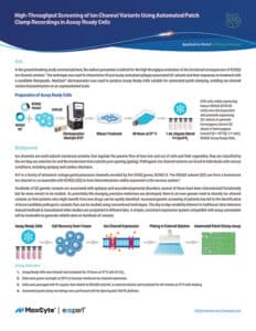
High-Throughput Screening of Ion Channel Variants Using Automated Patch Clamp Recordings in Assay-Ready Cells: In this study, researchers analyzed potassium channel variants of KCNQ2, which is associated with epilepsy. Using high-throughput screening assays, researchers characterized previously unstudied epilepsy-associated Q2 variants and their responses to treatment with a candidate therapeutic. MaxCyte electroporation helped generate assay-ready cells for automated patch clamping, enabling ion channel variant characterization on a then-unprecedented scale.
Presenter

Have more questions?
Send your question to one of our cell engineering experts.
Transcript
James Brady: Today we're going to be talking about MaxCyte’s scalable electroporation platform, and I'll be talking about how this technology can enable users to rapidly develop assays for screening ion channels, GPCRs and other drug targets.
I want to start with a brief discussion about cell-based assays, talk about why they're important, but also talk about some of the key challenges associated with developing a cell-based assay. And then I'm going to be talking about how MaxCyte’s electroporation platform helps you to address these challenges. It allows you to work with relevant cell types. You can screen multiple targets including complex multi-subunit ion channels. I'll give you some tips and tricks for optimizing your assays using MaxCyte electroporation. I'll highlight some of the key advantages of this platform: consistency as well as scalability of the transfection process, and also how you can cryopreserve cells without loss of function, so how you can essentially generate what we call assay-ready cells. And I'll walk through a brief case study about using assay-ready cells focusing on published data from the laboratory of Dr. Al George at Northwestern University.
The key advantage, or one of the big advantages of cell-based assays is it allows you to screen your drug targets in a physiologically relevant context working with disease-relevant cells and models. Of course, when you're working with primary cells or other physiologically relevant cells, those cells can be difficult to transfect. Other challenges with cell-based assays include the fact that you may be expressing complex multi-subunit targets, and often you have to optimize the stoichiometry of those subunits—that can be very challenging to do, particularly in the stable cell line. Many drug targets, particularly ion channels, can be toxic when they're over expressed. So in the case of stable cell lines, that toxicity is going to lead to either silencing or at least drift in expression levels every time you pass those cells.
Now, transfection could be used either to generate stable cell lines or for transient target expression. We have many MaxCyte customers using MaxCyte for stable cell line engineering, but today I'm really going to focus on transient target expression. The key advantages of transient transfection for assay development include the ability to express multiple variants or mutations without having to generate lots of different stable cell lines. Obviously, it is commonly used in basic research for target discovery pathway analysis, but in the case of MaxCyte, transient transfection can also allow you to do high-throughput screening with transiently transfected cells.
Just to give you a quick introduction to MaxCyte, Maxcyte’s been around for a little over 25 years, and our technology, or rather our company, was founded to commercialize a scalable electroporation technology, which we originally developed for clinical use, and clinical electroporation is still a very large part of our business. We take cells from patients, transfect them with receptors, chimeric antigen receptors, CARs, T cell receptors, CRISPR editing reagents, and give those back to patients to treat a variety of diseases. It has multiple advantages over viral vectors for gene delivery. But the point I want to make today is that over the years working with primary cells from patients, we've developed expertise working with very challenging cell types, and that expertise with challenging cells translates into benefits for customers that want to develop physiologically relevant cell-based assays.
So with MaxCyte, you get high efficiency with many cell types as well as high cell viability, you could load virtually any type of molecule and express a wide range of targets. I'll talk about how you can fine-tune your assay results and then, ultimately, how you can scale up that assay without having to reoptimize.
To start with a quick example of a customer that was working with a relevant cell type, in this case, we had a pharma company that was targeting CNS [central nervous system] diseases, and they wanted to screen their compounds in primary rat embryonic neurons. We first started off by optimizing transfection conditions using a GFP plasmid. Then we transfected in two of their drug targets, in this case labeled protein A and protein B. Cells were plated in 96 well plates post electroporation. You can see with just the GFP reporter, we could get about 40% to 50% of the cells showing robust GFP expression. You can also see normal neural morphology in these cells.
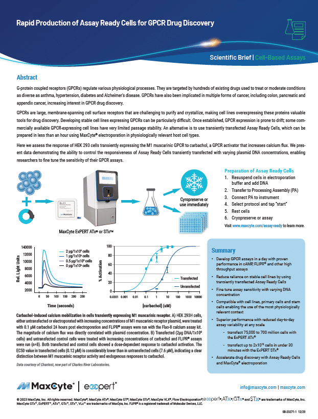
And then for their two proteins of interest, two points. First of all, you can see, in both cases, we got a robust assay response of their target proteins. Also, a point I want to keep coming back to is that we could dial in or modulate that assay response by changing the DNA concentration.
Here's another example of a customer working in physiologically relevant cells. In this case, the customer was screening for drugs targeting cystic fibrosis. So cystic fibrosis is caused by mutations in the CFTR protein. They wanted to develop a DI flux assay by co-transfecting yellow fluorescent protein, which acts as a HYlight sensor, along with various mutants of the CFTR. A lot of screening for CFTR is done in either A549 cells or fisher rat thyroid [FRT] cells. Neither of these cell lines is really ideal. Obviously, FRT is a rat cell line, not ideal for screening drugs targeting a human protein. And then while A549 is a human cell line, it's not a bronchial cell line.
CFP is a bronchial epithelial cell line. It's been challenging to generate stable cell lines expressing CFTR mutants. Here you can see, on the left, though that, they got very efficient transient expression of their yellow fluorescent protein. They could further optimize their assay response by varying the concentration of DNA for that YFP plasmid, and then also by further optimizing the ratio of the CFTR to the YFP plasmid, they could further dial in that assay response. At the bottom right, you can see their DI flux response comparing the two stable cell lines FRT and A549 with the transiently transfected CFBE cells.
And for this particular compound, they got comparable assay responses in all three cell types. Here's an example of a customer working with a complex target. This was a multi-subunit, voltage-gated calcium channel. For this particular assay, the customer transfected three different plasmids encoding three subunits of CAV2.2.
They actually transfected in a fourth plasmid, encoding an inward rectifying potassium channel. Then they screened these cells in a DI flex assay, a FLIPR assay. You can see a robust DI flex response on the transfected cells, also showing they could knock down that DI flux using conotoxin, which shows that these transfected cells were suitable for drug screening.
In this case, we had a customer that wanted to screen species-specific variants for TRIPV1. So rather than generating different stable cell lines for all the species that they're looking to screen, they found that MaxCyte transfection was a much faster approach. Here's a comparison of the rat versus the human TRIPV1 channel and their transiently transfected cells.
Three different DNA concentrations for each plasmid reused, all three DNA concentrations gave a robust assay response, and you can also see good Z prime scores for both their agonist and antagonist compound. So when you're trying to find the ideal species for in vivo studies, and you want to screen the different species specific isoforms in vitro, much faster to do transient transfection compared to making multiple stable cell lines for all the different isoforms.
Now in addition to varying the DNA concentrations, there are other ways to optimize your assay response with MaxCyte-transfected cells. In this case, I'm showing you data from an automated patch clamping assay, in this case for screening KV1.5. Again, the customer used two DNA concentrations to vary the assay response. And with a higher DNA concentration, they obviously had more cells expressing current. But you can say when they culture the cells at 37 degrees by 48 hours, fewer cells were expressing current. After culture at 37 degrees for extended periods of time, the assay response dropped off. But if they did a temperature shift and they cultured at 37 degrees and then shifted down to 28 degrees for another 24 hours, you could see almost a hundred percent of the cells were expressing current at that 48-hour time point.
Coming back to that point of optimizing the DNA concentration to optimize the assay response, I want to point out if you have a lot of different variants to screen, you don't have to redo that optimization step each time. To illustrate that point, these are customer data. Again, automated patch clamp data for screening voltage-gated potassium channels. Here they're looking at two different channels. One is KV1.3, in this case fused to GFP. So you can see on the left, the GFP allows you to visualize the high transfection efficiency, but here they're dialing in the DNA concentration with KV1.3. Somewhere between 80 to 100 micrograms per mil is the ideal concentration for KV1.5. Somewhere between 100 to 200 micrograms per mil works well, so the sweet spot for this particular class of channels and the cell line is probably about 100 micrograms per mil. So if you have the same cell line, the same assay, the same class of target, you don't have to keep redoing that optimization for all the different variants that you need to screen.
In addition to varying the DNA concentration, another way to dial in your assay response is to vary the plating density. So these are data from a publication from the Scripps [High-Throughput] Molecular Screening Center in Jupiter, Florida. In this case, they were developing an assay to screen for compounds targeting TRPM and this is another DI flex assay, a FLIPR assay. But here they've done a transfection and then they're plating the cells of 1536-well plates at different densities, anywhere from 500 or 4,000 cells per well. So in this case, 4,000 cells per well allowed them to maximize their assay window, as well as giving them good Z prime and good signal to background levels.
Now if you contrast this with, say, lipid-based transfection, typically with lipids you're doing a separate transfection in every well, so you get well-to-well variability in terms of transfection efficiency, but also lipids and other chemicals typically require you to keep your cells at a specific density during transfection, and that density may not be optimal for your assay response. With MaxCyte, you do the transfection and then you plate the cells, so you don't have that well-to-well variability for transfection efficiency, and you have the flexibility to plate your cells in any type of plate, any type of assay format.
And again, another key benefit of MaxCyte is the scalability. So the way we scale up the transfection process is we provide different consumables—we call them processing assemblies—that accommodate different cell numbers. At the small end of the spectrum, we have multi-well chambers. For example, you can see the R-50x8, the third consumable from the top, that has eight wells. Those hold about a million cells per well. So those are ideal for optimizing your DNA concentration or plasmid ratios.
Then you can scale up the process, uh, maybe using the R-1000 with 100 to 200 million cells to optimize plating conditions. Then when you've dialed in your DNA concentration, optimize the plating conditions, you can use Flow Electroporation®. The CL-2 shown at the bottom of the figure is the processing assembly we use for flow EP that can hold about 100 mils of concentrated cells, typically 10 to 20 billion cells. You mix in your plasmid DNA and then the cells are drawn into the electroporation chamber and three mil aliquot. So it's a repeated three ml transfection. We can process 10 to 20 billion cells in about 20 minutes, but you can see in the histograms on the left, these are K562 cells we transfected with GFP. Anywhere on that spectrum, you get the same transfection efficiency and there's no need to reoptimize your electroporation parameters or DNA concentrations as you scale up the transfection process.
And just to show how that scalability works for a real cell-based assay, these are customer-generated data. In this case, U2OS cells were co-transfected with a nuclear hormone receptor and a luciferase reporter plasmid. Here you can see they tested four different concentrations of that two-plasmid mixture. In this case, the highest DNA concentration gave them the best assay window. They took that same DNA concentration and went to Flow Electroporation with a billion cells.
You can see comparing a billion cells by Flow Electroporation versus 40 million cells in cuvette. Very consistent assay response. So again, no need to reoptimize as you go from that small scale to large scale. And then with that large-scale Flow Electroporation, you can generate enough cells either to plate for high-throughput screening, or those cells can be cryopreserved.
We don't place any restrictions on cryopreservation media. You can use DMSO, you can use commercial cryopreservation media. Cells can be frozen immediately after electroporation. Sometimes it helps to let them rest for an hour or so before freezing. Other customers like to plate the cells for a day or two and then cryopreserve the cells when their target is kind of at its maximum expression. And the advantage of that approach is that once you thaw those cells out, they're ready to load right away into your assay.
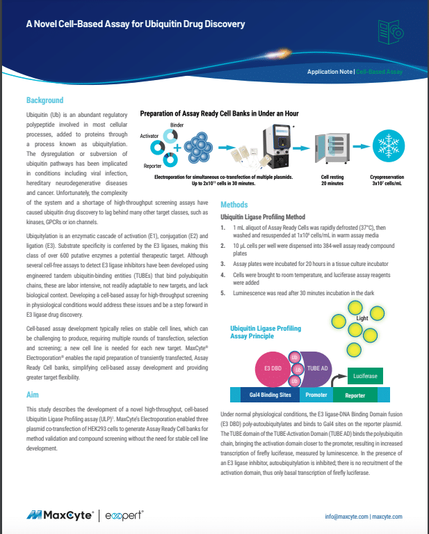
As an example of cryopreservation, here's more customer data. In this case, a customer, again, expressing a reporter plus a nuclear receptor in Jurkat cells. If you look at the luciferase response in freshly transfected cells versus cells that were transfected, frozen and thawed, you can see there's no loss of signal to background or assay response. And so again, the advantage of MaxCyte is you can do that small-scale optimization to figure out your plating conditions. DNA concentrations scale up that same transfection process using Flow Electroporation—you can generate typically up to 20 billion cells. Those can be plated and used immediately for high-throughput screening, or we can freeze them down to generate what we call assay-ready cells that can be thawed whenever it's convenient to run your assay.
I want to conclude with a quick case study of how one of our customers used MaxCyte to generate assay-ready cells. This is data taken from a publication from Dr. Al George's lab at Northwestern University. His lab is focused on potassium channel variants and their function in disease pathology. For this particular study, they were looking at variants of KCNQ2, which is associated with variance of this channel or associated with epilepsy.
KCNQ2 can form either homotetramers or it forms heterotetramers, but KCNQ3 and the disease phenotype is manifested in these heterotetramer. So what they did to develop these assays was they took a CHO cell line that was stably expressing KCNQ3, and they used MaxCyte to introduce KCNQ2 variants by transient expression. Forty-eight hours after transfection, they cryopreserved the cells and then those cells were thawed and they loaded them onto the SyncroPatch 768 for high-throughput patch clamping. And here's data they generated with 39 variants of KCNQ2, showing variant responses to their voltage step gradient.
Also, this particular study showed how these different variants responded to a compound used to treat epilepsy called retigabine. I don't really have time to do justice to this paper or this case study, but I do want to point out that on our website we have this case study as well as other drug-screening case studies that walk you through advantages of MaxCyte for cell-based assay development in more detail. I can also point you to many different publications from customers using MaxCyte for high-throughput screening and cell-based assay development.
To summarize again, highlighting key advantages of the MaxCyte platform for cell-based assay development: high transfection efficiency and viability with many cell types, including primary cells, physiologically relevant cells; the ability to co-transfect multiple plasmids, express complex targets; you can fine-tune that assay response by varying DNA concentrations, plasmid ratios, etc. The cells are healthy. Again, we develop expertise working with primary cells from patients. We have minimal impact on membrane biology. The cells are suitable for patch clamping, other types of assays. And obviously, large-scale transfection with MaxCyte is much faster than making stable cell lines, especially when you have multiple variants to screen; and the ability to cryopreserve those cells to generate assay-ready cells offers a further level of convenience and flexibility.
If people have questions or would like to see additional data, you can reach out to me directly. You can go to our website: We have many more case studies, publications and data that are available for a wide range of assays as well as other applications. Thank you very much for your attention.
